1-603-357-2455 — 686 Court Street Keene, NH 03431

TPLO Surgery

Cranial Cruciate Ligament (ACL) Tears and Your Pet’s Surgery

Torn Cranial Cruciate Ligaments (the equivalent of the “ACL”) in our canine friends are UNBELIEVABLY common, and it occurs for a different reason compared to humans.

  • Based on the dog’s INTERNAL knee biomechanics, they engage their ACL with each and every step that they take. This is in contrast to the human ACL which is engaged only when external force acts upon the joint.
  • Tears are most commonly caused by chronic “wear and tear”, compared to injuries that occur from traumatic events.
  • Risk of their other knee experiencing an injury to the cruciate ligament is ~50%.

Since we can see that the development of this problem in dogs is much more complex than in humans, and they experience different degrees of rupture (partial or complete), the canine condition is referred to as ‘cranial cruciate ligament disease’ (CrCLD). While the clinical signs (symptoms) associated with it can vary, the condition invariably causes rear limb dysfunction, lameness, and pain.

Diagnosing complete tears of the CrCL is easily accomplished using a combination of gait observations, physical examination findings, and radiography (X-rays).

Many treatment options are available for CrCLD. The first major decision is between surgical treatment and non-surgical (also termed conservative or medical) treatment/management. The best option for your pet depends on many factors, including their activity levels, size, age, skeletal conformation, and degree of knee instability. Deciding to pursue surgery is a big decision and we are here to help answer your questions and guide you to a choice that is right for your pet and your family.

We always recommend our patients see a Boarded Orthopedic Surgeon for a specialist opinion & repair; though if cost or distance are insurmountable obstacles, we are proud to be able to offer care at a local, General Practitioner level!

Understanding a bit more about the canine cruciate ligament

This is an X-ray (side view) of a dog’s normal knee:

Xray (side view) of dog’s normal knee:

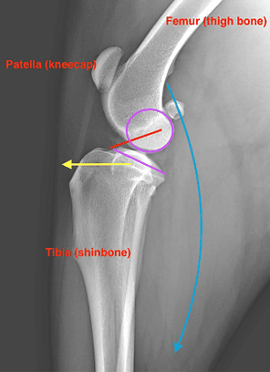
The ACL is labeled as the red line (Notice the trajectory/path that this ACL takes).

​​The joint surfaces are purple.

  • Notice how the bottom of the femur is round to permit flexion and extension of the joint on the flat part of the top of the tibia; however, the “flat” part is sloped (like a hill).
  • This also means that there is a shearing/slipping force that occurs during every step, as the “ball” is on a “sloped” hill.

The force that results from the calf muscles is shown as the blue arrow.

The primary force that the ACL counteracts during weight-bearing is shown as the yellow arrow… This force must be counteracted on EVERY step that your dog takes because of their biomechanics – The round joint surface of the femur slips off the sloped joint surface of the tibia as the muscles pull through the joint.

When the ACL is ruptured, the tibia (shinbone) slips forward relative to the femur. Your pet will likely experience joint effusion (swelling within the knee joint) resulting from inflammation. They may also experience early development of arthritis due to instability within the joint.

Understanding how the TPLO Procedure works

The cut in the bone “Osteotomy” is seen in red.

This is a side view (left) and front face (right) X-ray of a dog’s knee AFTER their TPLO procedure.

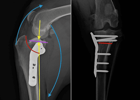
  • This is a semi-circular saw blade that allows the joint surface to be rotated (left image).
  • This cut is made parallel to the joint in the front face (“frontal”) plane (right image)

The joint surface (“plateau”) is seen in purple

  • The plateau is rotated so when your pet bears weight (yellow arrows) and their muscles engage (blue arrows) all the forces are transmitted through the joint and there is no abnormal shearing/sliding motion within their knee joint.

The metal bone plate and screws (bright white on the X-rays) are implanted to stabilize the bone in its new position until it has healed.

  • The bone takes approximately 8 weeks to heal, and during that time it is very important to not overload this implant.
  • Once the bone has healed, this implant is no longer active and is just there for the “free ride”.

General Rehabilitation After Surgery

The TPLO procedure requires 8 weeks of strict exercise restriction and physical rehabilitation. Our hospital will guide you through recovery with a week-by-week plan, ensuring the best possible outcome for your pet!

Educational information and images made possible through Veterinary Information Network and Veterinary Partner, with special thanks to our colleague Dr. Albert Lynch, DACVS-SA

Office Hours

Monday—Friday: 8:30 AM – 5:30 PM
Saturday:   Closed
Sunday:   Closed

Phone: 603-357-2455
Email:
[email protected]

686 Court Street Keene, NH 03431

Scroll to Top

Visit Our Online Pharmacy

Your pet’s food and medications delivered right to your door – with free shipping!*

*Orders over $49 and all AutoShip, RemindMe℠, and food orders. Free standard shipping within the continental U.S.